Spectral Doppler Settings Every Echo Student Should Know
Spectral Doppler lets sonographers measure blood flow velocity and direction. There are several adjustable settings that affect how clear and accurate that measurement is.
Velocity scale: This sets how the Doppler waveform is displayed. By convention, flow moving toward the transducer shows up above the baseline, and flow moving away shows below it (though this can be inverted). The scale should be adjusted so the waveform is as large as possible without aliasing — more on that below.
Sweep speed: This is how fast the waveform scrolls across the screen, typically set to 100 mm/sec as a default, ideally showing 2–3 waveforms (heartbeats) per screen for accurate measurement. For suspected cardiac tamponade physiology, breathing-related flow changes are easier to see with a slower sweep speed (around 25 mm/sec) because more waveforms (heartbeats) are visible at once across a longer period of time.
Sample volume size: This determines how large an area the machine is "listening" to for Doppler signal. Too large a sample volume creates a noisy, less precise signal. When the sample volume size is optimized, it gives a clean Doppler reading specific to the structure being evaluated.
Wall filters and gain: Wall filters remove low-velocity noise signals (clutter) so the actual flow signal is easier to see. Gain controls the overall brighteness of the Doppler signal; the goal is a smooth, clear waveform without being so amplified that noise drowns out the real signal.
The Echo Journal breaks down one echocardiography topic like this every Tuesday and Thursday for over 9,000 sonographers and cardiologists worldwide.
The 3 Types of Spectral Doppler
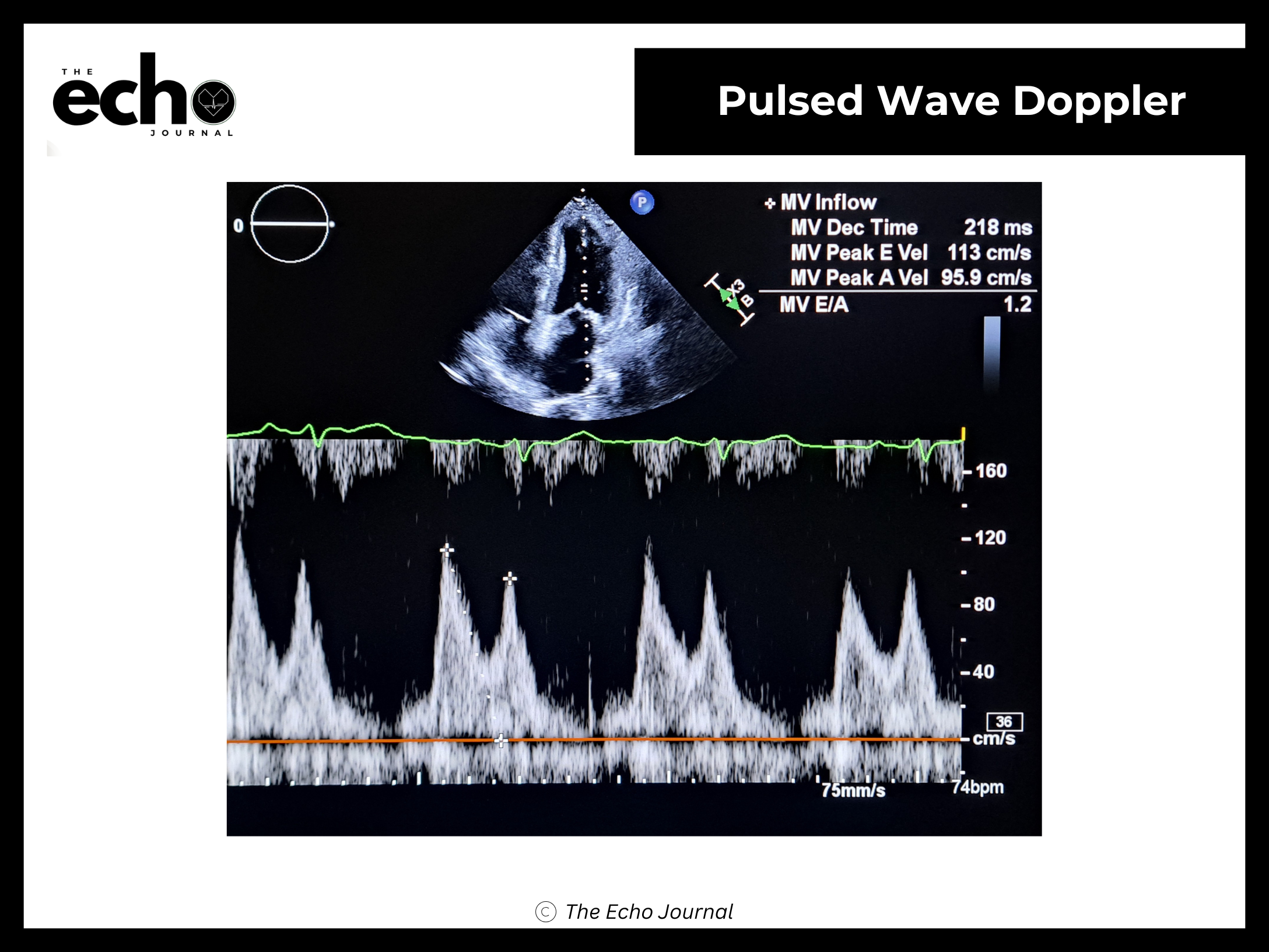
Pulsed-wave (PW) Doppler: Used to measure blood flow velocity at a specific depth. Its main limitation is aliasing — when blood is moving too fast for the system to accurately display, the waveform appears to "wrap around," making it look inaccurate. This happens when the actual velocity exceeds half of what's called the pulse repetition frequency, also known as the Nyquist limit. Pulsed wave Doppler waveforms have what is called a “spectral window”. The spectral window on pulsed-wave Doppler is the clear, hollow, or open area located underneath the systolic or diastolic spectral waveform curve. In a healthy vessel with normal laminar flow, red blood cells move at a uniform speed, leaving this clean, dark space below the velocity tracing.
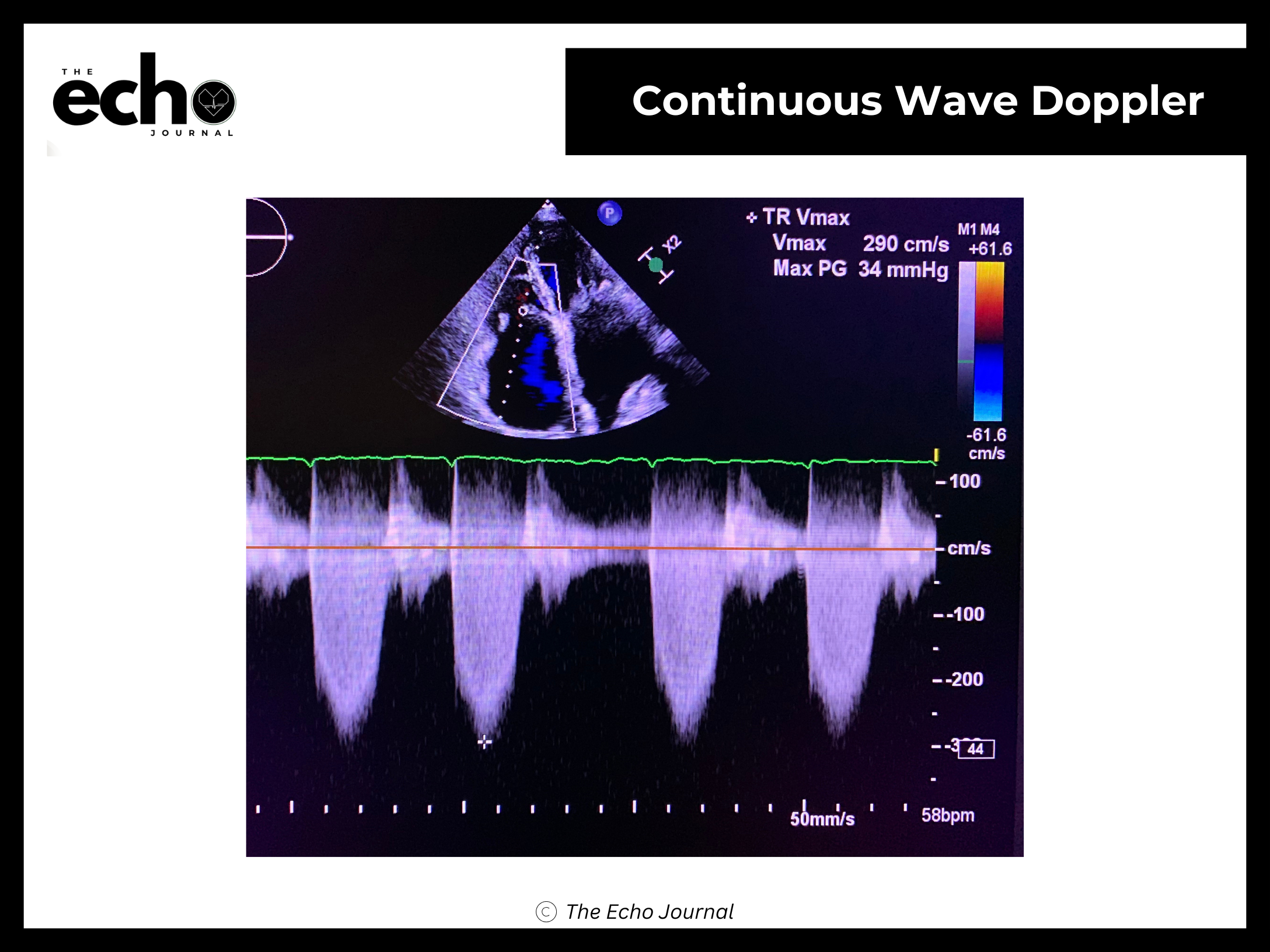
Continuous-wave (CW) Doppler: Used for measuring high velocities with no aliasing limit, since the machine is continuously transmitting and receiving rather than pulsing. The tradeoff here is that CW Doppler can't tell you exactly where along the beam path a signal originated because it captures everything along that line.
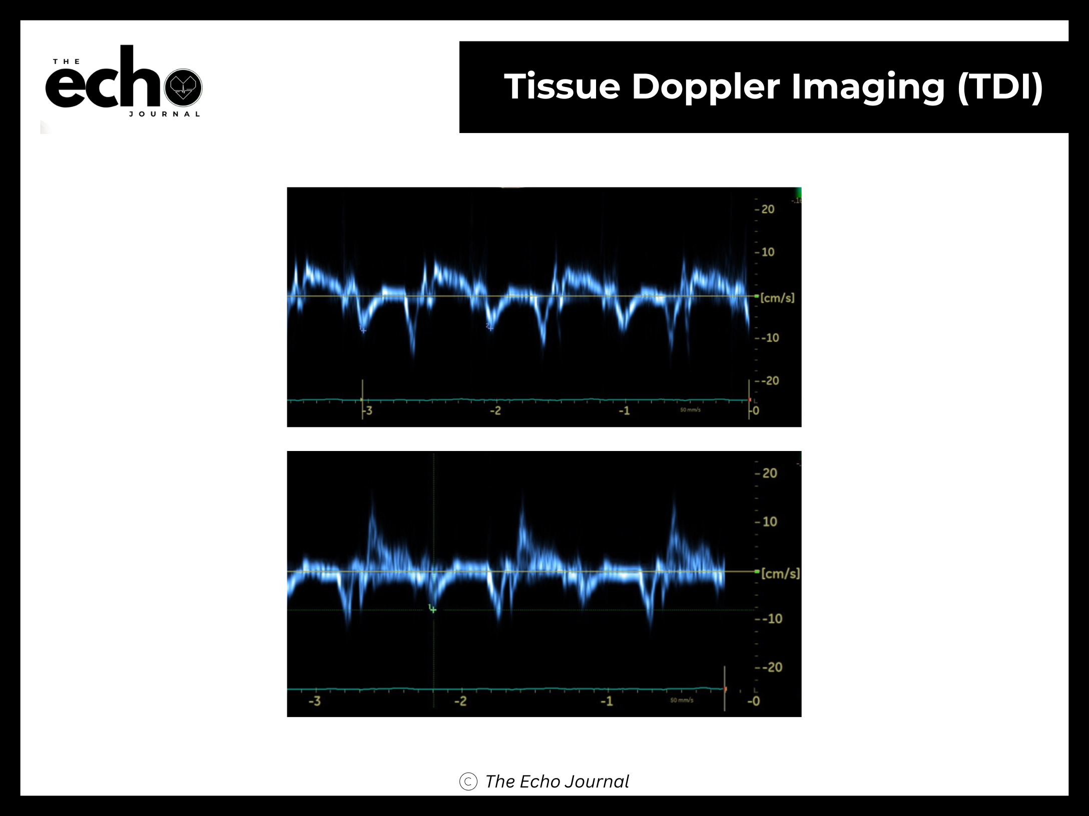
Tissue Doppler imaging (TDI): A specialized Doppler mode used to measure the movement of heart muscle itself (rather than blood), commonly at the mitral and tricuspid valve annulus. It detects much slower velocities than blood-flow Doppler, so it requires different filter and gain settings. It measures the displacement of the annulus towards the cardiac apex during systole.
High-pulse repetition frequency (HPRF) Doppler: Used when PW Doppler hits aliasing limits. It increases the number of sample volumes being measured, which raises the maximum measurable velocity, but introduces a tradeoff called range ambiguity, where it becomes harder to tell exactly which sample volume a signal is coming from.