How to Get an Apical 3 Chamber View on Echocardiography
Position the patient in the left lateral decubitus position, rolled fully onto their left side, all the way onto the hip, with the left arm raised and tucked under or behind the head. This isn't optional comfort positioning; rolling the patient brings the heart forward against the chest wall and shifts lung tissue out of the beam's path.
From the A2C view, rotate another 60 degrees counterclockwise. The aortic valve and outflow tract come into view.
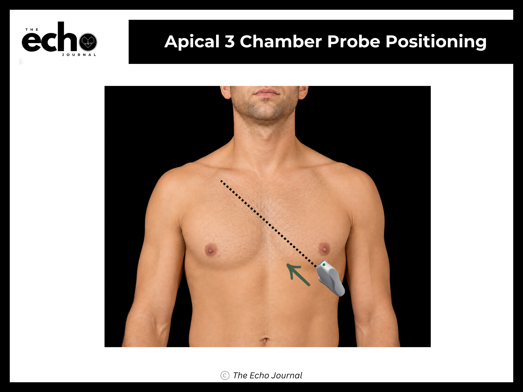
Structures you should be able to identify:
Left ventricle (LV) — anteroseptal and posterior walls
Left atrium (LA)
Mitral valve (MV)
Left ventricular outflow tract (LVOT)
Aortic valve (AV) and proximal aorta
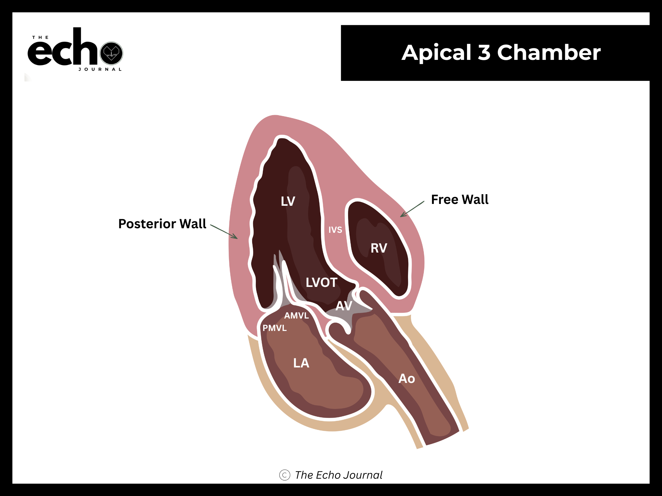
Getting it right: this view shows the same structures as PLAX, but from the apex. That matters because it gives you a beam angle nearly parallel to LVOT flow, which is exactly what you need for accurate Doppler measurements.
The Echo Journal breaks down one echocardiography topic like this every Tuesday and Thursday for over 9,000 sonographers and cardiologists worldwide.