How to Get Apical 2 Chamber View on Echocardiography
Position the patient in the left lateral decubitus position, rolled fully onto their left side, all the way onto the hip, with the left arm raised and tucked under or behind the head. This isn't optional comfort positioning; rolling the patient brings the heart forward against the chest wall and shifts lung tissue out of the beam's path.
From the A4C view, keep the probe in exactly the same spot and rotate approximately 60 degrees counterclockwise. The right-sided chambers drop out of the image and you are left with the LV and LA only.
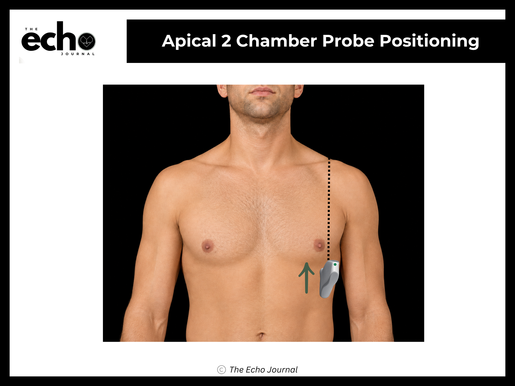
Structures you should be able to identify:
Left ventricle (LV) — anterior and inferior walls
Mitral valve (MV) — each mitral leaflet is divided into 3 scallops, anterior (A1,A2,A3) and posterior (P1,P2,P3), some seen in this view
Left atrium (LA)
Getting it right: you know you have a true A2C when no right ventricle is visible anywhere in the sector. If you can still see a sliver of RV, keep rotating.
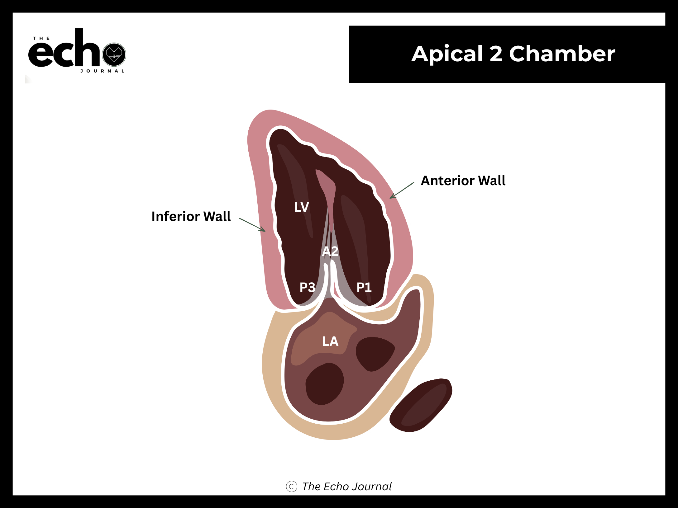
Getting it right: you know you have a true A2C when no right ventricle is visible anywhere in the sector. If you can still see a sliver of RV, keep rotating.
The Echo Journal breaks down one echocardiography topic like this every Tuesday and Thursday for over 9,000 sonographers and cardiologists worldwide.