How to Get an Apical 5 Chamber View on Echocardiography
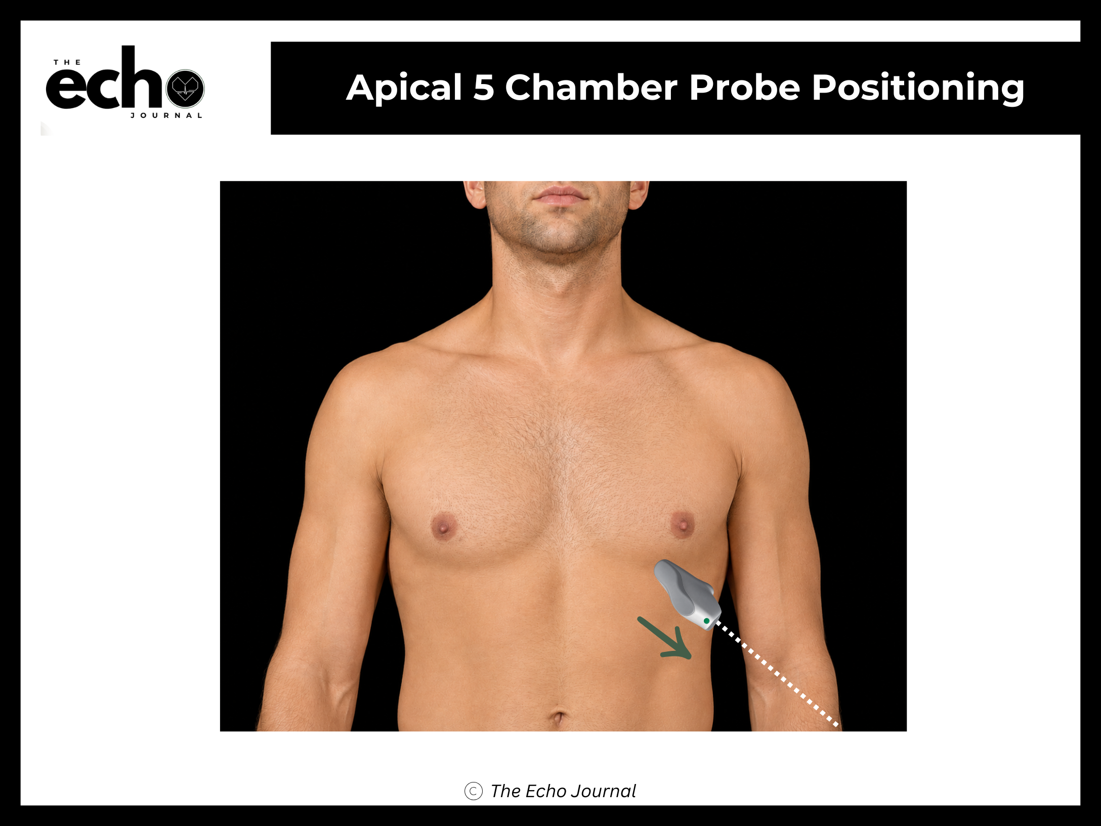
Position the patient in the left lateral decubitus position, rolled fully onto their left side, all the way onto the hip, with the left arm raised and tucked under or behind the head. This isn't optional comfort positioning; rolling the patient brings the heart forward against the chest wall and shifts lung tissue out of the beam's path.
From the A4C view, keep the probe in the same spot and rotate to the index marker to 4 o’clock position. Tilt the beam anteriorly, the tail of the probe drops toward the bed, aiming the beam slightly more toward the patient's chest wall. The left ventricular outflow tract and aortic root swing into the center of the image, between the two atria.
Structures you should be able to identify:
Left ventricle (LV) and left atrium (LA)
Right ventricle (RV) and right atrium (RA)
Mitral valve (MV)
Interventricular septum (IVS)
Left ventricular outflow tract (LVOT)
Aortic valve and aortic root (Ao) — the "fifth chamber" the view is named for
The Echo Journal breaks down one echocardiography topic like this every Tuesday and Thursday for over 9,000 sonographers and cardiologists worldwide.
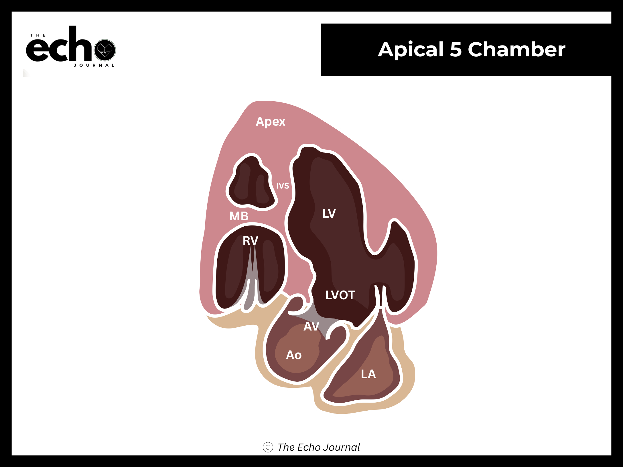
Why this view matters: the LVOT sits nearly parallel to your beam here, which is exactly the alignment Doppler requires. This is the view used for pulsed-wave Doppler at the LVOT for stroke volume and cardiac output, and for continuous-wave Doppler across the aortic valve when assessing aortic stenosis. Every one of those measurements is angle-dependent, so a sloppy A5C produces a confidently wrong number.