How to Get the Apical 4 Chamber View on Echocardiography
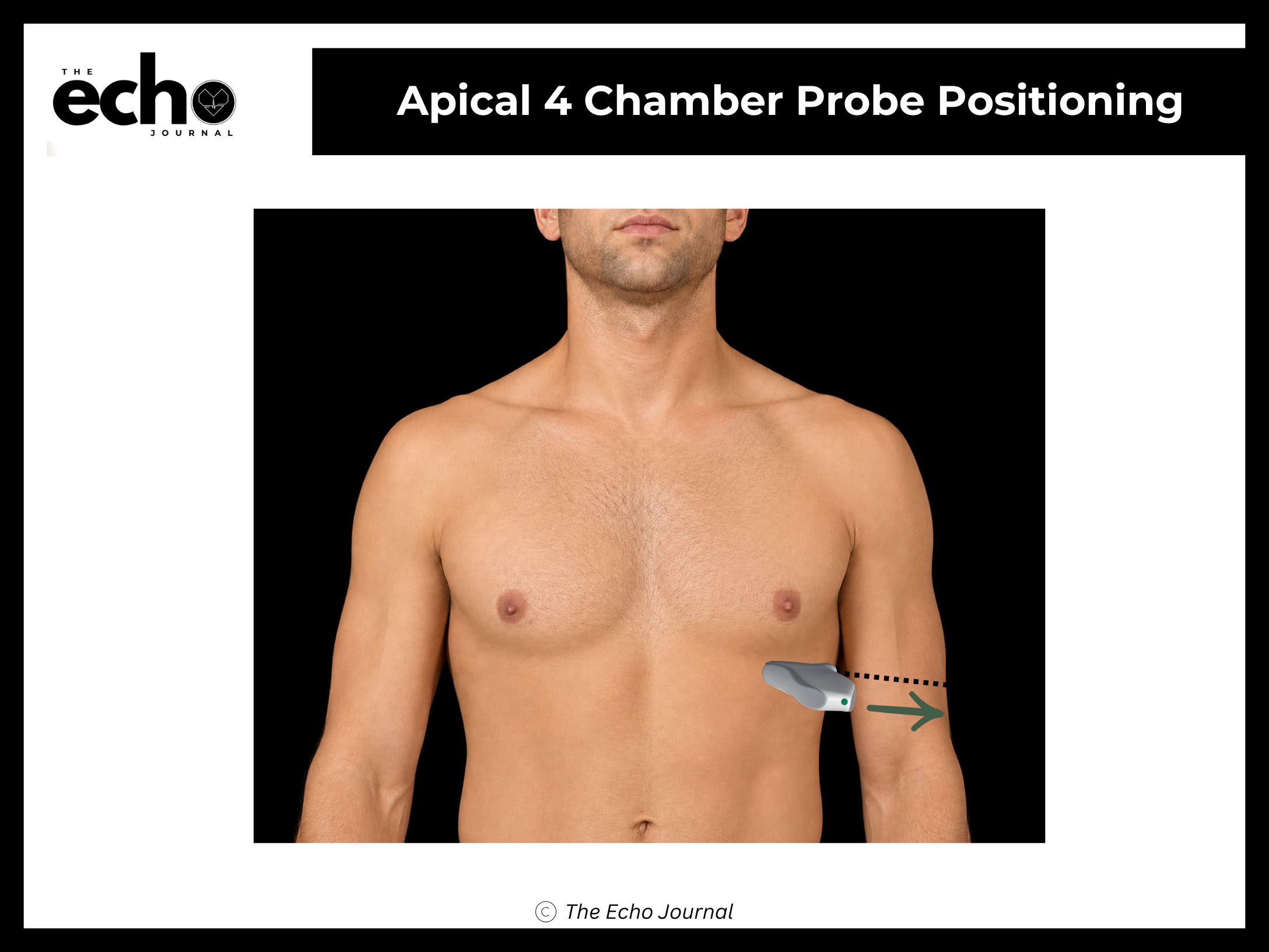
Position the patient in the left lateral decubitus position, rolled fully onto their left side, all the way onto the hip, with the left arm raised and tucked under or behind the head. This isn't optional comfort positioning; rolling the patient brings the heart forward against the chest wall and shifts lung tissue out of the beam's path.
Place the phased array transducer below the left breast at the apical impulse — the spot where you can feel the heartbeat tapping against the chest wall. This marks the cardiac apex, the tip of the heart, which points down and toward the patient's left. Point the index marker toward the patient's left side (roughly the 3 o'clock position) and aim the beam up toward the patient's right shoulder.
Structures you should be able to identify:
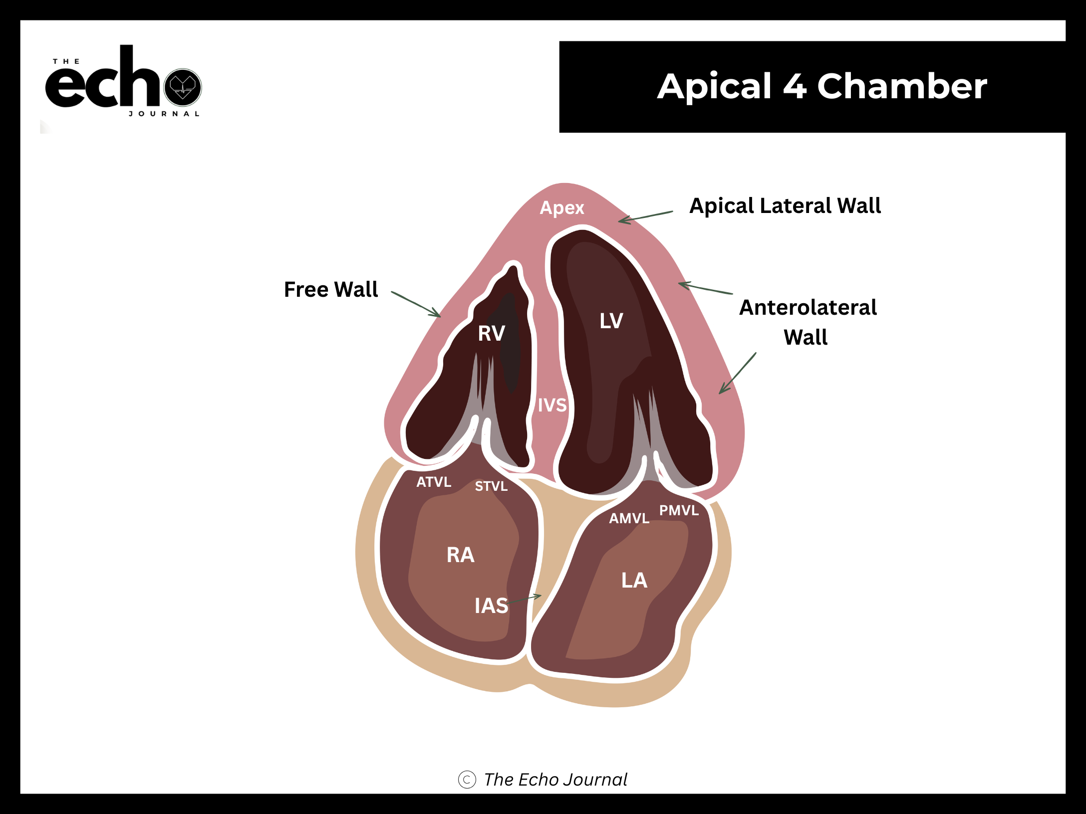
Left ventricle (LV), left atrium (LA) and apex
Right ventricle (RV) and right atrium (RA)
Mitral valve (anterior and posterior leaflets) and tricuspid valve (anterior and septal leaflets)
Interventricular septum (IVS)
Interatrial septum (IAS)
The Echo Journal breaks down one echocardiography topic like this every Tuesday and Thursday for over 9,000 sonographers and cardiologists worldwide.
Getting it right: the number one beginner error here is foreshortening — cutting across the apex instead of through it, which makes the LV look shorter and rounder than it really is and falsely inflates ejection fraction. If the apex looks blunt or thick, you are too high on the chest. Move down an interspace and more laterally, not just angle differently.