How to Get the Right Ventricular Outflow Tract (RVOT) Long Axis View on Echocardiography
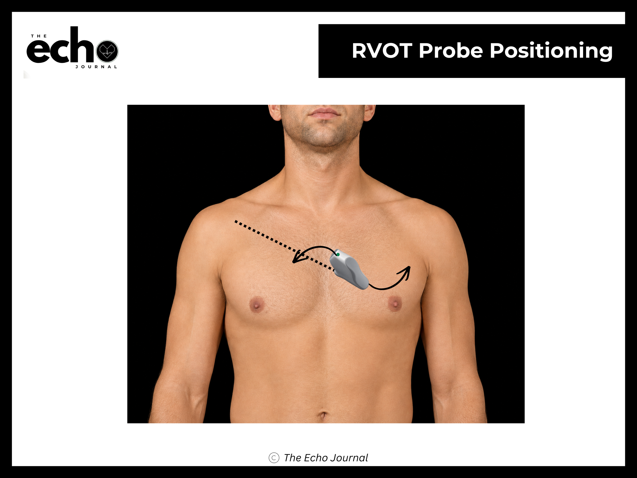
Position the patient in the left lateral decubitus position, rolled fully onto their left side, all the way onto the hip, with the left arm raised and tucked under or behind the head. This isn't optional comfort positioning; rolling the patient brings the heart forward against the chest wall and shifts lung tissue out of the beam's path.
From the PLAX view, again keep the index marker toward the patient's right shoulder at roughly 10 o'clock and do not rotate. This time tilt the beam superolaterally, aiming the beam up and toward the patient's left shoulder, the opposite direction from the RV inflow view. The tail of the probe drops toward the patient's right hip.
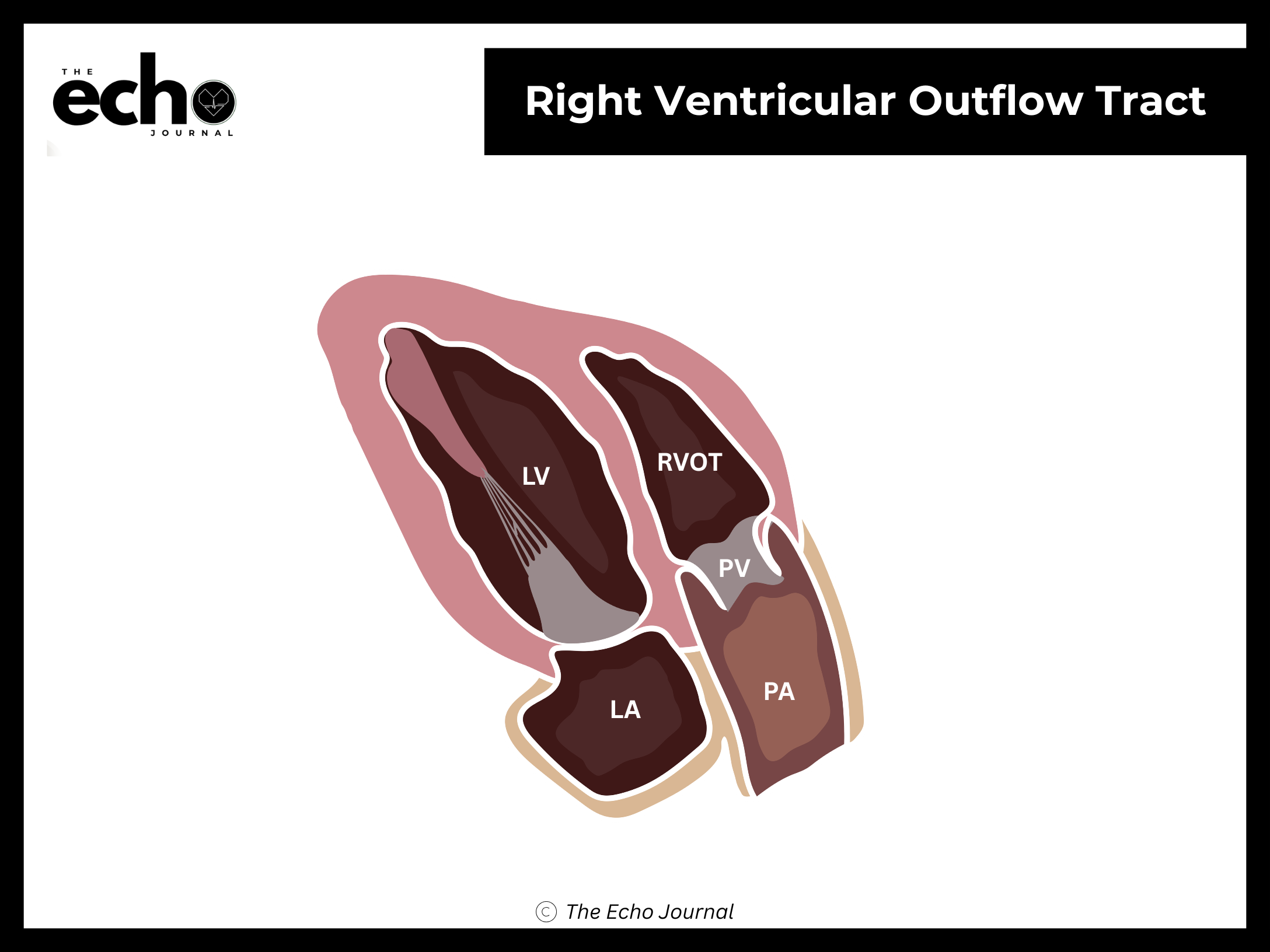
Structures you should be able to identify:
Right ventricular outflow tract (RVOT)
Pulmonic valve (PV)
Main pulmonary artery (MPA)
A portion of the left ventricle and aortic root, at the edge of the sector
Why this view matters: it gives you a beam angle nearly parallel to flow through the RVOT and pulmonic valve. That's the alignment you need for RVOT velocity time integral, for pulmonic stenosis gradients, and for the pulmonic regurgitation jet used to estimate pulmonary artery diastolic pressure. As with the LVOT, all of these numbers are angle-dependent.
The Echo Journal breaks down one echocardiography topic like this every Tuesday and Thursday for over 9,000 sonographers and cardiologists worldwide.
Getting it right: the pulmonic valve and main pulmonary artery should run roughly along the axis of your beam, not across it. And don't confuse this with the short-axis great vessel view, which also shows the RVOT and pulmonic valve, that one requires a 90° rotation, while this view comes from tilting alone. Same distinction as the A5C versus apical long axis: tilting changes the slice, rotating changes the plane.
A useful way to hold these two in your head: the PLAX position is the hub, and the two right-sided views are opposite tilts from it. Tilt down and medial toward the right hip for inflow. Tilt up and lateral toward the left shoulder for outflow. One probe position, two opposite angles, the entire right heart.