How to Get the Parasternal Short Axis Aortic Valve Level on Echocardiography
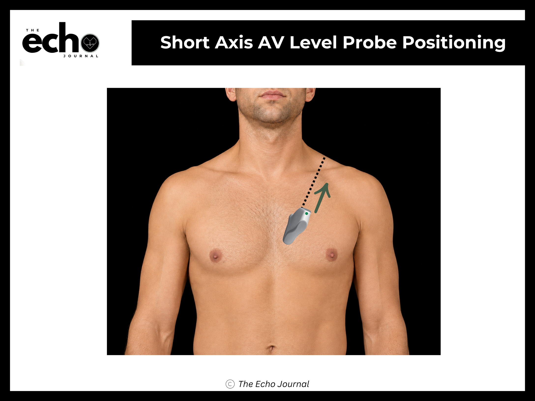
Position the patient in the left lateral decubitus position, rolled fully onto their left side, all the way onto the hip, with the left arm raised and tucked under or behind the head. This isn't optional comfort positioning; rolling the patient brings the heart forward against the chest wall and shifts lung tissue out of the beam's path.
Start in the parasternal long axis, with the index marker pointing toward the patient's right shoulder, at roughly 10 o'clock. Without sliding the probe off that spot, rotate it 90° clockwise so the marker now points toward the patient's left shoulder, at roughly 1–2 o'clock. That single rotation lands you at the aortic valve level, and the marker stays at 1–2 o'clock for the entire short-axis sweep, you will not rotate again.
Structures you should be able to identify:
Aortic valve (AV) — the three cusps in the center, forming the classic "Mercedes-Benz" sign when closed
Right-coronary cusp (RCC)
Non-coronary cusp (NCC)
Left-coronary cusp (LCC)
Right atrium (RA)
Tricuspid valve (TV)
Right Ventricle (RV) and the right ventricular free wall are seen in this view
Right ventricular outflow tract (RVOT), wrapping around the aorta
Pulmonic valve (PV) and main pulmonary artery
Left atrium (LA)
Interatrial septum (IAS)
The Echo Journal breaks down one echocardiography topic like this every Tuesday and Thursday for over 9,000 sonographers and cardiologists worldwide.
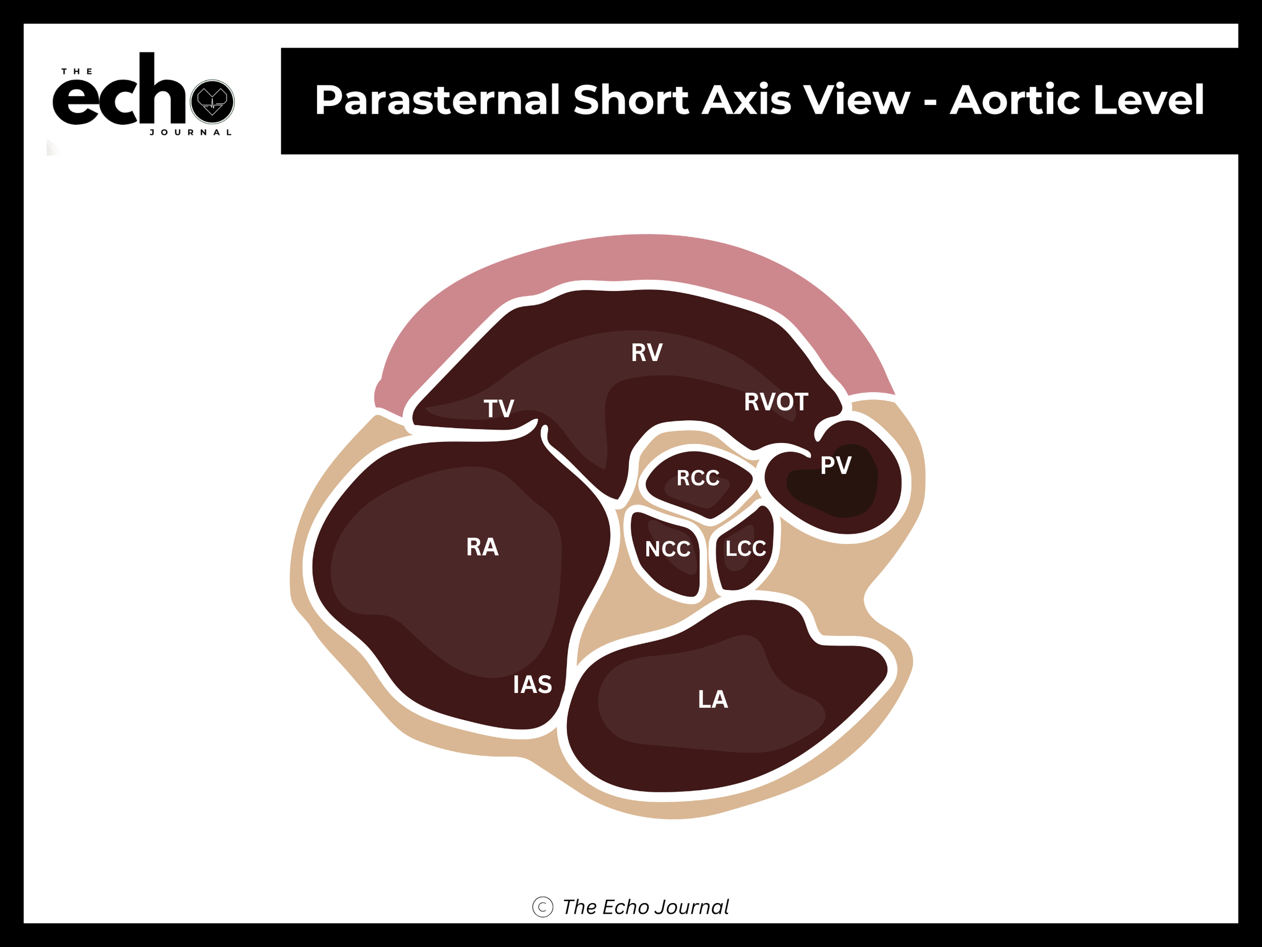
Getting it right: small tilting adjustments matter more than big ones here. You want all three aortic cusps in the same plane at the same time. If you only see two cusps clearly, you're cutting the valve obliquely.