How to Get the Right Ventricular Inflow (RVIT) View on Echocardiography
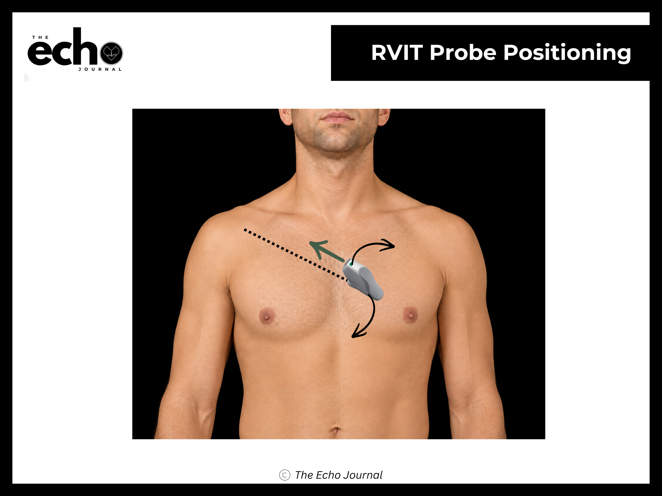
Position the patient in the left lateral decubitus position, rolled fully onto their left side, all the way onto the hip, with the left arm raised and tucked under or behind the head. This isn't optional comfort positioning; rolling the patient brings the heart forward against the chest wall and shifts lung tissue out of the beam's path.
From the PLAX view, keep the index marker where it is, toward the patient's right shoulder, at roughly 10 o'clock, and do not rotate. Tilt the beam inferomedially, aiming down and toward the patient's right hip. The tail of the probe comes toward the patient's left shoulder. The left-sided structures drop out of the image and the right atrium, tricuspid valve, and right ventricle fill the sector.
Structures you should be able to identify:
Right atrium (RA)
Right ventricle (RV) — the inflow portion and the RV free wall
Tricuspid valve (TV) — typically the anterior leaflet superiorly and the septal or posterior leaflet inferiorly, depending on your exact angulation
Coronary sinus, entering the RA near the tricuspid annulus
Inferior vena cava (IVC) and the Eustachian valve, at the RA border
Why this view matters: the tricuspid regurgitation jet often aligns better with your beam here than in any apical view, which makes this the view that rescues an RVSP estimate when the apical window is poor. It's also your best look at the RV free wall and at TV leaflet anatomy in a long-axis plane.
The Echo Journal breaks down one echocardiography topic like this every Tuesday and Thursday for over 9,000 sonographers and cardiologists worldwide.
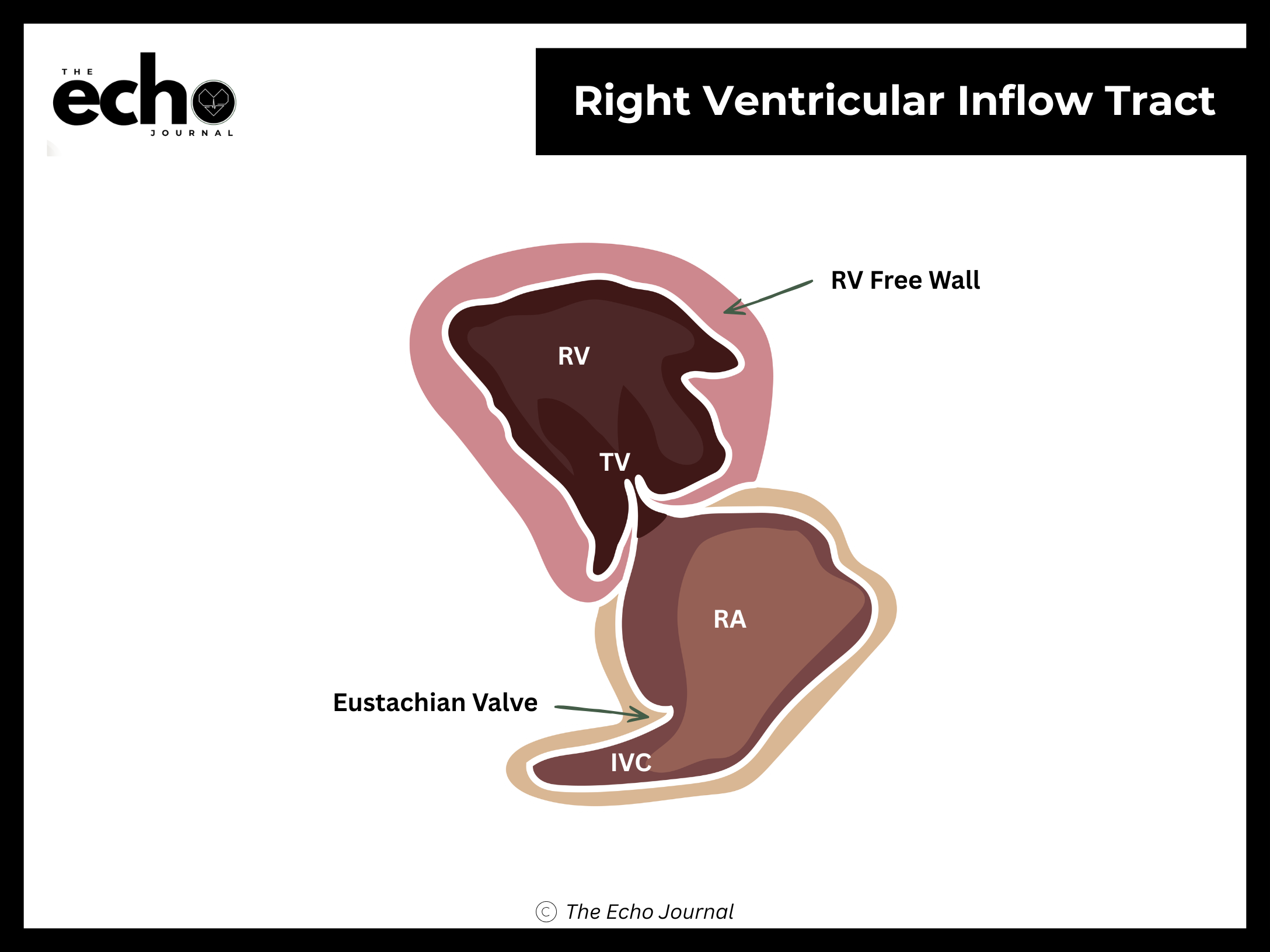
Getting it right: if any left ventricle is still visible in the sector, you haven't tilted far enough — a true RV inflow view contains no LV at all. You may need to slide down one interspace as you tilt, since the RV inflow sits lower on the chest than the PLAX plane. Sweep gently once you're there; the alignment that gives you the best TR jet is often a degree or two off the alignment that gives you the prettiest 2D image.