How to Get the Parasternal Short Axis Left Ventricle on Echocardiography (Basal, Mid, Apical)
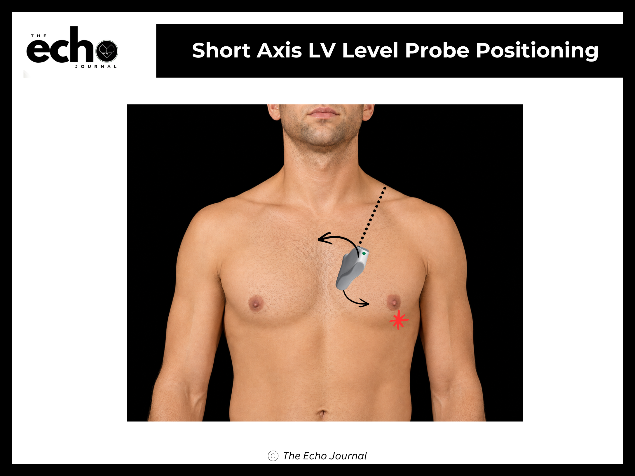
Position the patient in the left lateral decubitus position, rolled fully onto their left side, all the way onto the hip, with the left arm raised and tucked under or behind the head. This isn't optional comfort positioning; rolling the patient brings the heart forward against the chest wall and shifts lung tissue out of the beam's path.
Start in the parasternal long axis, with the index marker pointing toward the patient's right shoulder, at roughly 10 o'clock. Without sliding the probe off that spot, rotate it 90° clockwise so the marker now points toward the patient's left shoulder, at roughly 1–2 o'clock. That single rotation lands you at the aortic valve level, and the marker stays at 1–2 o'clock for the entire short-axis sweep. You will not rotate again.
From there, tilt the beam inferolaterally, toward the LV apex — marked by the asterisk in the image above. In practice, the tail of the probe rises toward the patient's right shoulder while the beam sweeps down and out toward the left hip. Each degree of tilt walks you down the ventricle to the next level.
There are three levels to capture, in this order:
1. Mitral valve level — the anterior and posterior mitral leaflets open and close in the center of the LV cavity, producing the "fish mouth" appearance. Structures: RV, IVS, anterior mitral leaflet (AMVL), posterior mitral leaflet (PMVL), LV
2. Papillary muscle level — both papillary muscles are visible inside a round LV cavity. This is the level used for assessing regional wall motion and LV shape. Structures: RV, IVS, anterolateral papillary muscle (ALPap), posteromedial papillary muscle (PMPap), LV
3. Apical level — a small, thick-walled circle with no papillary muscles in view. Structures: LV apex
The Echo Journal breaks down one echocardiography topic like this every Tuesday and Thursday for over 9,000 sonographers and cardiologists worldwide.
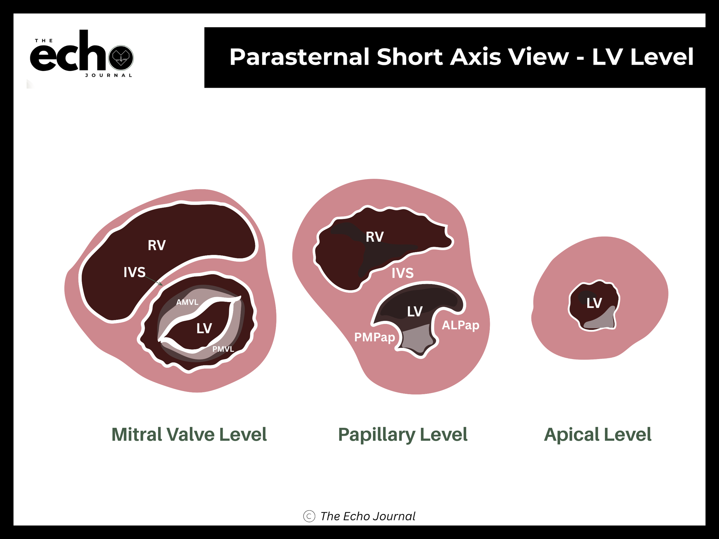
Getting it right: as you approach the apex, tilting alone often isn't enough: the true apex sits lower and more lateral on the chest than you expect. Slide the probe laterally, down toward the apical cap, rather than tilting to the point that your image becomes an oblique, oval-shaped slice. A round LV means you're on axis. An oval LV means you're cutting through it at an angle and any measurement you take will be wrong.