How to Get the Parasternal Long Axis (PLAX) View on Echocardiography
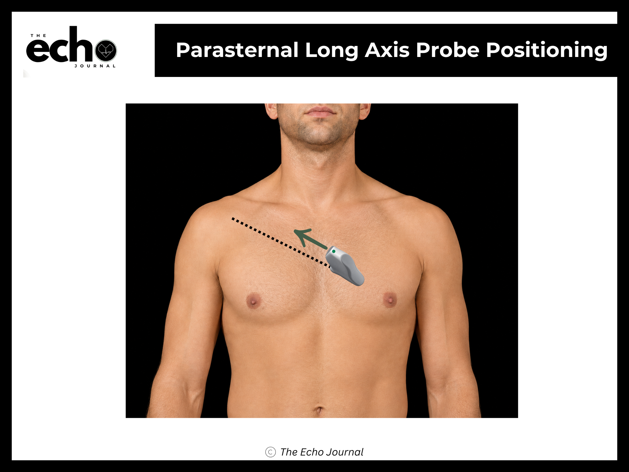
Position the patient in the left lateral decubitus position, rolled fully onto their left side, all the way onto the hip, with the left arm raised and tucked under or behind the head. This isn't optional comfort positioning; rolling the patient brings the heart forward against the chest wall and shifts lung tissue out of the beam's path.
Place a phased array transducer in the 3rd or 4th left intercostal space, immediately beside the sternum. Point the index marker toward the patient's right shoulder (roughly 10 o'clock). The beam should cut through the long axis of the left ventricle.
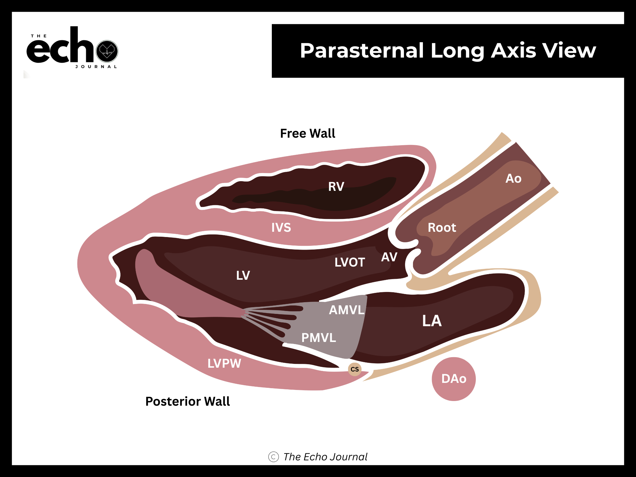
Structures you should be able to identify:
Right ventricle (RV) — the chamber closest to the probe, at the top of the image
Left ventricle (LV)
Interventricular septum (IVS)
Posterior left ventricular wall (LVPW)
Left ventricular outflow tract (LVOT)
Aortic valve (AV) and aortic root
Mitral valve (MV) — both the anterior (AMVL) and posterior leaflets (PMVL)
Left atrium (LA)
Descending thoracic aorta (DAo), seen in cross-section behind the LA
Coronary sinus (CS), can also be seen in this view and is relevant when evaluating for persistent left superior vena
Pericardium, as a bright line below the posterior LV wall and around the entire right ventricle in this view
The Echo Journal breaks down one echocardiography topic like this every Tuesday and Thursday for over 9,000 sonographers and cardiologists worldwide.
Getting it right: the goal is a true long axis, which means the beam is perpendicular to the left ventricle and you are seeing the widest possible LV cavity. If the LV looks small or the apex in bouncing into the picture, you're off axis and may need to rotate, slide, or tilt the probe.